Glaucoma

Glaucoma

Going from the Laymans explanation to the scientific is helpful in understanding what is involved in attempting to wrestle this Progressive Optic Neuropathy into a manageable submission time and again. The break down in management comes by patient non-compliance to care directives as Glaucoma is virtually symptomless in the more common Open Angle format such that patients often   questions the expense of treatment and examination over time and the corneal irritation and occasional  systemic side effects to such Rx’d drugs.

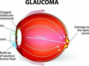
True, Glaucoma does involve the resultant manifestations of too high a pressure within an eye, but this is an over simplification however, as other vascular diseases can increase Glaucomas likelihood and certainly deteriorating health can exasperate this disease even if Intra Ocular pressure seems to be at a pre determined Targeted level therefore this family of diseases the Glaucomas are multi factorial; for the most part of an Inherited Template with 2 identified Genes in the Trabecular meshwork to date. No one is ever cured of Glaucoma but rather managed. Literature would have you know it is the second leading cause of blindness in the US having only the occasional report of blurred vision and the occasional coloured ring around lights even in the Open Angle form. Contrasted to Macular Degeneration that involves the Retinal elements devoted to fine discriminate vision and relating to the central 20 degrees of vision: Glaucoma involves for the largest part of its disease course the retinal tissue that provides the non- discriminate sight. We know that with the disease the first changes occur in the mid-periphery of vision but at the same time a sizable amount of vision loss has occurred before a person realizes they are forming tunnel vision.
Glaucoma

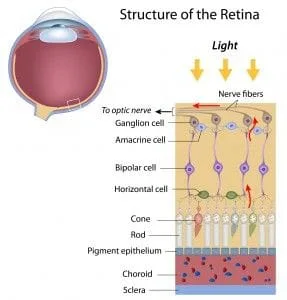
If you run an IOP consistently over 21 mm hg this will give use reason for concern but at the same time if you do not have any demonstrated Parametric field loss or documentable Optic Nerve Change, correct you do not by definition have Glaucoma at this point but rather Ocular Hypertension (OH). It’s here where we arrive at this grey area (O.H.) poorly communicated and poorly understood by patients who are reluctant to return for multiple testing. At present in Canada the go to confirmer of any very early Optic nerve head changes would be by running an HRT over a 3 visit 18 month course by still many O.M.D’s or by employing an O.C.T by OD’s of record of ownership. Apparently,.Only 3 % of O.D.’s have O.C.T’s. Here in either case we are looking for an interval or indices change over successive tests (progression). At the same time the astute clinician could use comparative stereoscopic digitized photos and direct observation with a biomicroscope and hand held magnification. I must add now that even though some Glaucoma Clinicians stay rooted to the HRT stating they have such a large data base of comparatives to refer too. The OCT has now sufficient database over all ethnic groups to be a better glaucoma tool as it has the ability to detect the loss of ganglion cell components under the macula where 50% of the Ganglion cells exist.

As an aside Data bases are not restricted in Canada in the same way they are in the U.S. by the F.D.A. so OCT’s here have expanded data bases. Macular Ganglion Cell Thinning as a entity gives us the ability to predict glaucoma 3 to 4 years sooner than the HRT. At least this is my experience to date. The Oct seems to indicate Glaucoma 11 to 13 years sooner than other means. A further addition is there is now the Designations PG and PPG. Parametric Glaucoma where a field change occurs and Pre – Parametric where O.N.F.L., optic nerve fiber layer thinning and a loss of Ganglion cells under the macula proceeds any Field or (Parametric Change).

Back to the biomicroscope and digital image method though still useful at this point in order for the next clinician to reach the same conclusion they would run a H.R.T. and expect a similar resultant course. The Humphrey Field Analyzer run annually in this case tests viewing points out to 24 or 30 degrees of the central retina. A cautionary note however is if a defect shows up here it has been determined that 35 to 40% of the fibers have already been damaged if we are talking about pressure only Glaucoma once we factor out fiber loss do to age and omit cardiovascular involvement also.

So if Glaucoma has been determined and a target pressure of low teens has been achieved on going you must still test for progressive loss of nerve fibers. Undetected pressure spikes and blood flow dynamics can alter the disease course. The normal range for IOP is between 11 and 21 mm hg 15.5 being an average.

What does confound many a patient is that they can be told that they have an IOP (intra ocular pressure)) below 20 and at the same time has Glaucoma. This would be considered to be Normal Tension Glaucoma, yes included in the Open Angle Glaucoma family but with a differing etiology. Apparently 61% of Primary Open Angle Glaucoma patients present with a mean pressure below 21 mm Hg. (Optometry Rounds volume 1,issue 4, 2013.)

The held belief is that something is affecting the perfusion pressure to the Optic nerve such that it will be damaged such that a demonstrated field loss will show in the results of parametric, (Field Testing) eventually. The clue- in, to the clinician is changes in Optic nerve head appearance in the early going, possible pressure spikes in serial tonometry: a contact lens called the Trigger fish is coming for this use, isolated disc hemorrhages (‘Drance Hemorrhages’ Lasting up to 3 months) and most importantly confirmable sequenced interval changes on successive HRT tests and now of course the High Def O.C.T.
With Normal Tension Glaucoma it is believed the person’s blood pressure drops too low at portions of their sleep cycle (Nocturnal Hypotension) or that they have Vasospasms like with Raynauds Disease, or that they have some underlying adverse cardiovascular disease  that would contribute to vasoconstriction. Highest incidence of N.T.G.Japanese Females.What we are really speaking to hear is the ocular perfusion dynamics to the optic nerve(mean ocular perfusion pressure). New methods are en route to track this .It has been postulated that the short posterior cilliary arteries may collapse inward during sleep in certain individuals as the body achieves a supine position. Decreased systemic blood perfusion in the presence of increased I.O.P. can adversely affect retention of retinal nerve fiber. Certain individuals classified as having Nocturnal Hypotension will lose large amounts of nerve fiber if they receive Night time B.P medication and Persons suspected of NTG should be questioned about Snoring and Sleep Apnea also.. Apnea meaning the person stops breathing during their sleep cycle. Eating heavy within 2 hrs of sleep, heavy alcohol and /or coffee ingestion and being as little as 10 pounds over weight contributes to sleep apnea. Sleeping on ones back contributes to it .A person should drink enough water to keep passages hydrated. This relates to less serious presentations. In A 1999 Bern Switzerland study of 114 OSA white subjects. Obstructive sleep apnea pts yielded Glaucoma in 7.2% of that population where as the accepted incidence in a non-OSA white population is 2%. The the M.O.A. is not totally agreed upon but it appears to be the eventual Hypoxic episodes that kill nerve fiber ( Specifically ganglion cells ). Whether IOP spikes or not is not totally agreed upon. A longitudinal Taiwanese Study of 6,072 subjects 1,012 with sleep apnea and 5,060 without had an occurrence of 11.26 per 1000 persons in the sleep apnea group and 6.76 in the non sleep apnea group a therefore 1.67 times greater risk factor but not the sole factor as determine 5 years out. ( Ophthalmology 2013 ). Non the less Nightly IOP Prostaglandin med should be given when a OCT reveal loss of Retinal Nerve Fiber before Parametric field loss, especially when fiber loss has reached a important level and the damage is progressing. As stated before perfusion pressure while be more commonly measured by pending instrumentation of greater use ie the O.R.A.; The Ocular Response Analyzer and the Pascal Tonometer; these will correlate well. For now utilizing the B.P’s and I.O.P. in a lengthy formula Ocular perfusion can be estimated such that caution can be noted to review or rethink proposed meds   when the number falls below 50 (see Milton and Thomas). Precise Formula for mean opp=2/3{DBP+1/3( SBP – DBP)} -IOP. Opp means ocular perfusion pressure. These persons warrant also review at a Sleep Clinic. The procedure is called Polysomography. Maxiofacial Surgeons are the persons who might alter the interior structure of your throat to aid breathing. One out of five people with sleep apnea have mild presentation, one out of fifteen have moderate yet to have Thirty or more episodes in a 7 hr night of sleep is a severe problem as is a 60 to 90 sec cessation of breathing. This info from the Obstructive Sleep Apnea (O.S.A.) Institute. To leave off there is controversy about sleep apnea and the impact it has on optic nerves; STILL. Pachymetry is the process of measuring the central thickness of an individuals cornea with an ultrasonography device Why someone would do this at least once to all new patients is to find persons with a corneal thickness under 555 nanometers. This number is the central standard measurement. You also will discover persons who have corneal ectatic disease like Keratoconus a stromal corneal dystrophy which might be slowed or possibly arrested with collagen cross linking treatment very early; the verdict is mixed here. African Americans typically run 520 to 540 nanometers considered thin corneas while Caucasions run 580 to 600 considered thick. Know this very startling fact if you read a 24 pressure on a thick cornea that reading is Falsly High and more like really 20 or 21 mm hg. But if you read that same 24 mm hg on a thin cornea you have a problem going forward as that True Pressure could reasonably be 27 or 28 mm hg. There are differing views as to the correctness of such an assertion none the less it warrents consideration. Different Optic Nerves can handle differing pressures before they demonstrate verifiable damage. KNOW THIS FACT! Persons with Normal Tension Glaucoma tend to have thin corneas and often large optic nerves. All this information is courtesy of the Ocular Hypertension Treatment Study. What this now better relates to is something Called Hysteresis; the Biomechanics of the stated cornea and how this interrelates to Glaucoma’s progression. Hysteresis can be measured with a highly sophisticated machine called the Ocular Response Analyzer. The current finding is if you have a low corneal Hysteresis and you are put on Glaucoma Med you will get a greater lowering in intraocular pressure with that same med versus had that persons hysteresis value had been higher. Say a 30% lowering versus a 18% lowering. Apparently corneal hysteresis as a relative value can forecast the integrity of the optic nerve structure and how it may respond to drug treatment. ( Latest Academy of Ophthalmology findings). Just to touch on Hysteresis what is evaluated is” the dampening Viscosity “of a Cornea. Apparently a raised I.O.P. will cause distention of the optic nerves laminar cribotic matrix.

This will falter with prolonged raised I.O.P. insult and fibers will die. The Cornea with a higher measured Hysteresis value serves a better damper to this strain and because the drug can raise the Hysteresis value it in turn must assist the laminar cribosa matrix slowing what is termed atopsis or fiber melt structural compromise .In these 2 new directives with the Ocular Response analyzer you seem to be dealing more with the Mechanical Theory of Glaucoma and with the Sweapt Source O.C.T. and blood flow tracking at the nerve head the Vascular Theory of Glaucoma.Personally I think they play off each other and are therefore a combined entity.

NOW THE STATS. With regards to Glaucoma just like Diabetes a large portion of the population do not know they have it and just like Diabetes damage can occur before a concrete diagnosis is placed. With Glaucoma it is referred as the “Thief of Sight” as it is never going to give you a clue unlike Diabetes until you are really engaged. Really Shocking Facts: at 65 years of age you are 7 times more likely to have Glaucoma than under that age. Certain ethnic groups approach a near 20% incidence at or around age 75, Hispanic, African American (The Glaucoma research Institute and the Latino eye study). Incidence 1 to 2 % over 40 and about 10% at 70 years of age (UK data). Luckly only 15% go blind. To high by my view. According to a British Study 10% become eventually non responsive to drug therapy and require surgery. Hopefully the ROCK family of drugs and better understanding of blood flow dynamics will reduce the need for surgery as it is not free of complications such as Kissing Choroidals.

Again remember IOP is not the only operative here. Hopefully better monitoring and control measures will reduce this ie: a vaccine as an adjunct treatment. If you have a family member with Glaucoma your risk rose 10 times to 10% if its your sibling who has glaucoma and 4 times to 4% if your mom or dad has or had it. With a Provincial Government in the Province of B.C. that is Clevearly hiding the Rationing of Preventative Diagnostic Eyecare Services by alluding serious Eye Disease only effects those over 60 so they suggest  that this segments eyecare is best relgeted to spectacle shops with trumped up auto refractors think again folks. Yes it may be fact that Glaucoma measured with current measures has a realization of 1 % in persons younger than 45 years old. Don’t back away from the fact that a genetic tests once devised  could redefine this variable and also take note that from 45 to 60 years of age the incidence jumps by 31% to that under age 45.Questioning now the plausibility that 80% of the people with glaucoma are over 60 years of age and there is the rapid jump after 40? If you chose to buy the Governments argument you will completely ignore the fact that Diabetes is on the rise in Canada attributable to Dietary Choice and a rapidly developing sedentary lifestyle. Know that if you are Myopic to any degree

Irrespective of age your risk for Glaucoma went up. They use to say that Diabetes seemed to give you a 1.8 times greater realization for Glaucoma but Now according to John’s Hopkins and the work done by the Centers for Excellence this statement is INCORRECT. Diabetes is not a risk factor; in fact it may be Neuroprotective by the release of Anti Vegs. Non the less Diabetes is a rapidly expanding biggy of the 4 vision loses CNIB (Canadian National Institute for the Blind) looks at.

When you draw over the entire age Paradim 20 to 70 years of age Diabetic Retinopathy leads as the source of potential vision loss. But it is the leading cause of Vision Loss under 50. According to the C.N.I.B.Of visitations to the CNIB Glaucoma ranks 7.6% and is ascribed a conservative incidence of < 2 %.Half the people that have Glaucoma do not know it.

Locations

Find us on the map

Hours of Operation

Our Regular Schedule

Harbourside Optometry Clinic

Monday:

9am – 5:30pm

Tuesday:

9am – 5:30pm

Wednesday:

9am – 5:30pm

Thursday:

9am – 5:30pm

Friday:

9am – 5:30pm

Saturday:

By Appt.

Sunday:

Closed

Beach House Optometry Clinic

Monday:

9:00 am-5:30 pm

Tuesday:

9:00 am-5:30 pm

Wednesday:

9:00 am-5:30 pm

Thursday:

9:00 am-5:30 pm

Friday:

9:00 am-5:30 pm

Saturday:

Appointment Upon Request

Sunday:

Closed

Testimonials

Reviews From Our Satisfied Patients

  • "Patient Testimonials: Coming Soon..."